Pages

Thursday, November 17, 2011

Awake Craniotomy

History:-

26 yr old gentleman admitted with c/c of:-

  • Right focal seizures-14 months

Examination:-

  • No neurological deficits

Investigations:-

  • MRI brain showed left frontal hypointense lesion with mild enhancement with perifocal edema in premotor cortex


Treatment:-

  • Awake craniotomt with gross total excision done under local anasthesia and sedation

Post-operative period:-

  • No neurological deficits

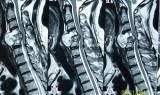
Post-operative CT scan head


Recurrent Chondrosarcoma

HISTORY :
  • 53 yr old gentleman operated earlier for low grade chonrosarcoma in 2005
  • Received post-operative radiation therapy in same year
  • Asymptomatic till Aug. 2011
  • Pain right C5 distribution since 2 months

EXAMINATION:

  • Grossly intact neurological status
  • MRI Cervical Spine:
  • C4-C6 extradural lesion with cord compression


CT angiography neck vessels:
  • Left vertebral artery block

TREATMENT:

  • Tumor decompression with C4-C6 corpectomies with C3-C7 fusion with expandable cage done

Histopathology:

  • Well diffrentiated chondrosarcoma

Postoperative X-Ray:


Hypothalamic hamartoma


HISTORY :
  • 11 yr old boy admitted with c/c of :-
  • Gelastic seizures-6 years, frequency-1/day, no relief inspite of trial of multiple antiepileptics
  • Intellectual impairment-6 years
EXAMINATION :

  • Impaired intellectual performance
  • No neurological deficits

INVESTIGATIONS:

TREATMENT:

  • Tumor excised thorough transcranial route

POST-OPERATIVE PERIOD:

  • No further episodes of seizures

Tuesday, November 1, 2011

4th Ventricular Tumor

5 yr old girl child admitted with c/c
  • Walking difficulty-2 years
  • Speech difficulty-1 year
  • Incoordination in the hands-6 months
EXAMINATION:-
  • Conscious, awake
  • Dysarthric speech
  • B/l cerebellar signs with gait ataxia
INVESTIGATIONS:-
  • MRI brain


TREATMENT:-
  • Suboocipital craniectomy with gross total excision done
HISTOPATHOLOGY:-
  • Pilocytic Astrocytoma

Hypothalamic Tumor

37 year old gentleman admitted with c/c of
  • Left hemicranial headache-3 months
  • Drooping of left eyelid-2 months

EXAMINATION:-

  • Left partial ptosis
  • Diplopia on seeing left, down and outward

INVESTIGATIONS:-

  • CT Scan head was normal
  • MRI brain-T2 & FLAIR showed hyperintense lesion in the hypothalamic area


TREATMENT:-
  • Fusion of CT and MRI brain done and coordinates calcucated
  • Biopsy done stereotactically